
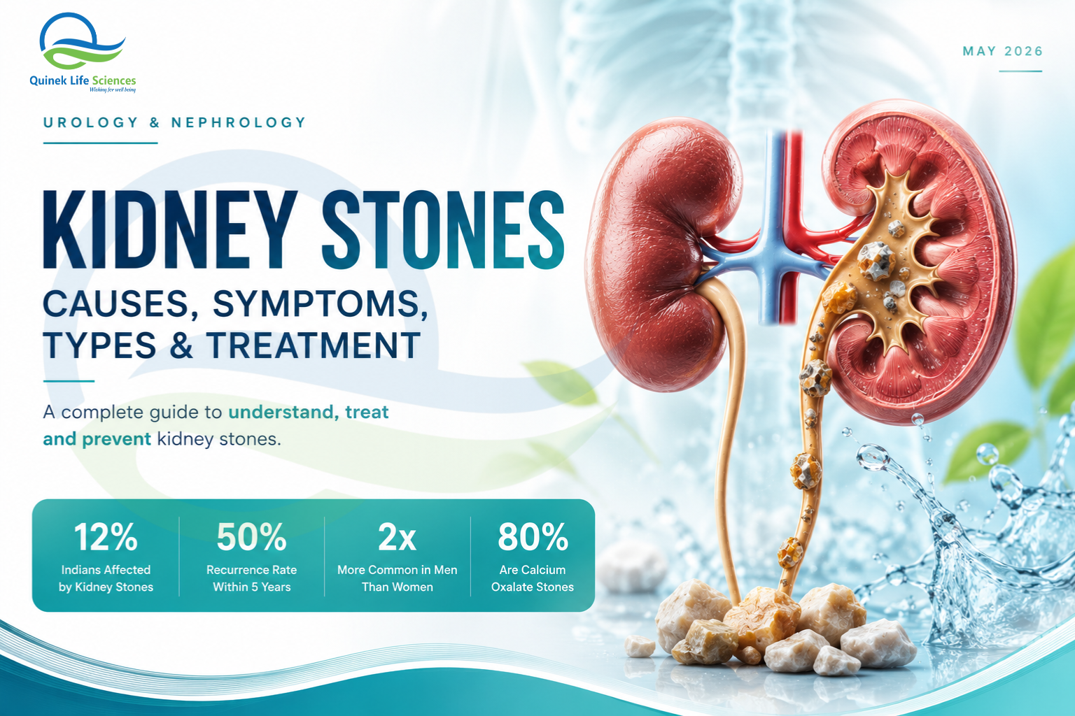
There are few experiences in medicine as viscerally described as renal colic — the pain of a kidney stone passing through the ureter. Patients describe it as the worst pain of their lives. It comes in waves, offers no comfortable position, and can last for hours. It sends hundreds of thousands of people to emergency departments across India every year.
And yet, for a condition this common and this painful, kidney stones are remarkably preventable. Most stones form due to identifiable metabolic, dietary and hydration factors that can be addressed. Most stones recur — but the recurrence is not inevitable. The problem is that most patients who pass a stone are told to “drink more water” and sent home, without a metabolic workup, without dietary guidance and without a prevention plan. Five years later, many of them are back in the same emergency department with another stone.
This article explains kidney stones comprehensively — for patients who want to understand what happened and how to prevent it happening again, for general practitioners managing the acute presentation, and for urologists and nephrologists looking for a complete clinical reference.
IN THIS ARTICLE
- What Are Kidney Stones?
- Types of Kidney Stones — Why It Matters
- How Kidney Stones Form
- Symptoms — From Silent to Severe
- Risk Factors — Who Gets Kidney Stones?
- How Kidney Stones Are Diagnosed
- Treatment — From Pain Relief to Stone Removal
- Prevention — The Most Important Section
- Frequently Asked Questions
- Quinek Life Sciences — Urology & Nephrology Support
What Are Kidney Stones?
Kidney stones — medically termed renal calculi or nephrolithiasis — are solid mineral and salt deposits that form inside the kidneys when urine becomes supersaturated with certain substances. When the concentration of stone-forming minerals (calcium, oxalate, uric acid, phosphate) exceeds the urine’s capacity to keep them dissolved, crystals begin to form and aggregate into stones.
Stones can range from less than a millimetre (the size of a grain of sand) to several centimetres (large enough to fill the renal pelvis — called a staghorn calculus). Small stones may pass spontaneously through the urinary tract with little or no symptoms. Larger stones can obstruct urine flow, cause severe pain, lead to urinary tract infections and, if left untreated, cause kidney damage.
KIDNEY STONES IN INDIA — KEY DATA
Prevalence of kidney stones in India: approximately 12% of the population · Source: NCBI — Nephrolithiasis in India
India is part of the global “stone belt” — a region with particularly high stone prevalence due to climate, diet and water quality factors
The hot, arid northern and western states (Rajasthan, Gujarat, Punjab, Haryana) have among the highest kidney stone prevalence in India
Recurrence rate without preventive intervention: up to 50% within 5 years
Types of Kidney Stones — Why It Matters
The type of kidney stone determines the underlying metabolic cause, guides dietary advice and dictates the most appropriate prevention strategy. Stone analysis — of a passed or surgically retrieved stone — is one of the most important and most underperformed investigations in clinical practice.
How Kidney Stones Form
Stone formation is fundamentally a problem of urine chemistry — when the balance between stone-forming substances and stone-inhibiting substances tips in the wrong direction. Understanding this chemistry is the key to understanding why stones form and how to prevent them.
Supersaturation
When urine becomes too concentrated — from dehydration, low fluid intake or high dietary mineral load — stone-forming ions exceed their solubility threshold and begin to crystallise.
Low Citrate
Citrate is the kidney’s primary natural stone inhibitor — it binds calcium and prevents crystal aggregation. Low urine citrate (from high protein diet, metabolic acidosis or chronic diarrhoea) dramatically increases stone risk.
Urine pH Extremes
Persistently acidic urine favours uric acid stone formation. Persistently alkaline urine favours calcium phosphate and struvite stone formation. Urine pH is a key therapeutic target.
Anatomical Factors
Urinary stasis from structural abnormalities (horseshoe kidney, medullary sponge kidney, ureteropelvic junction obstruction) creates conditions where crystals can aggregate without being flushed away.
Symptoms — From Silent to Severe
Kidney stones do not always cause symptoms. Small stones lodged in the kidney without obstructing urine flow can remain completely asymptomatic for years and are discovered incidentally on imaging done for another reason. It is when a stone moves into the ureter and begins to obstruct urine flow that the classic and dramatic presentation of renal colic begins.
EMERGENCY — WHEN TO SEEK IMMEDIATE CARE
Fever + flank pain + obstructing stone = urological emergency. Do not wait. An infected obstructed kidney can progress to urosepsis and life-threatening septic shock within hours. Emergency ureteral stenting or percutaneous nephrostomy is required to drain the obstruction before definitive stone treatment.
Risk Factors — Who Gets Kidney Stones?
How Kidney Stones Are Diagnosed
CT KUB — The Gold Standard
Non-contrast CT of the kidneys, ureters and bladder (CT KUB) is the gold standard for diagnosing kidney stones — with a sensitivity of over 95% and the ability to detect all stone types, including radiolucent uric acid stones invisible on plain X-ray. It confirms stone size, location and degree of obstruction — all of which guide treatment decisions. Reference: NCBI — CT in Diagnosis of Urolithiasis
Ultrasound
Ultrasound is the preferred first-line imaging in pregnancy, children and for serial monitoring — avoiding radiation exposure. It is highly sensitive for stones in the kidney and for hydronephrosis (ureteral obstruction), but less sensitive for ureteral stones, particularly in the mid-ureter.
Urine Analysis & Culture
Dipstick and microscopy for haematuria, pyuria and crystalluria. Urine culture to exclude infection — critical before any stone intervention. 24-hour urine collection (for calcium, oxalate, citrate, uric acid, pH, volume) is the cornerstone of metabolic workup for recurrent stone formers.
Blood Tests
Serum calcium, phosphate, uric acid, creatinine, electrolytes and parathyroid hormone (PTH). These exclude underlying metabolic causes — hyperparathyroidism, gout, renal tubular acidosis — that must be identified and treated to prevent recurrence.
Treatment — From Pain Relief to Stone Removal
Acute Pain Management
NSAIDs (diclofenac, ketorolac) are the first-line analgesics for renal colic — they are superior to opioids in multiple trials for pain control and have a direct anti-spasmodic effect on ureteral smooth muscle. Antiemetics (ondansetron, metoclopramide) are added for nausea. IV fluids if the patient is vomiting or dehydrated.
Medical Expulsive Therapy (MET)
Alpha-blockers (tamsulosin) relax ureteral smooth muscle and significantly increase the spontaneous passage rate of stones up to 10mm in size. They are recommended as adjunctive therapy for distal ureteral stones being managed conservatively. Reference: NEJM — Medical Expulsive Therapy for Ureteral Stones
Watchful Waiting
Stones ≤5mm pass spontaneously in 70–98% of cases. Conservative management with adequate hydration, analgesia and MET — with follow-up imaging to confirm passage.
ESWL
Extracorporeal shock wave lithotripsy — non-invasive sound wave fragmentation of stones up to 2cm. Effective for renal and proximal ureteral stones. Multiple sessions may be needed.
Ureteroscopy (URS)
Flexible or rigid ureteroscopy with laser lithotripsy — highly effective for ureteral stones of all sizes and renal stones up to 2cm. The most commonly performed stone intervention in India.
PCNL
Percutaneous nephrolithotomy — for large (>2cm), complex or staghorn calculi. Requires general anaesthesia, a percutaneous access tract into the kidney. Gold standard for large stone burden.
Prevention — The Most Important Section
Stone removal treats the stone. Prevention treats the disease. Without a prevention strategy, up to 50% of patients will form another stone within 5 years. Prevention is not complicated — but it requires understanding the specific stone type and the patient’s individual urine chemistry.
Frequently Asked Questions
Q. How long does it take to pass a kidney stone?
It depends on stone size. Stones smaller than 4mm pass spontaneously in approximately 80% of cases, usually within 1–2 weeks. Stones 4–6mm pass in about 60% of cases, often taking 2–4 weeks. Stones larger than 6mm rarely pass without intervention. Stone location (upper, mid or distal ureter) also affects passage time — distal ureteral stones pass most readily.
Q. Should I restrict calcium in my diet if I have calcium stones?
Counterintuitively, no. Restricting dietary calcium actually increases the risk of calcium oxalate stones — because without sufficient calcium in the gut to bind to dietary oxalate, more oxalate is absorbed and excreted in the urine. Maintain normal dietary calcium (dairy and calcium-rich foods) while moderating oxalate intake. This is one of the most important — and most commonly misunderstood — dietary points in stone management.
Q. Does drinking lemon juice help prevent kidney stones?
Yes — lemon juice is a practical, evidence-supported source of dietary citrate, which is the kidney’s primary natural stone inhibitor. Lemonade therapy (diluted fresh lemon juice) has been shown to increase urinary citrate and reduce stone recurrence in some studies. It is a useful adjunct to, but not a replacement for, adequate fluid intake and specific medical therapy.
Q. Can kidney stones damage the kidneys permanently?
Yes — if left untreated, obstructing kidney stones can cause hydronephrosis (kidney swelling from backed-up urine) and, over time, irreversible renal parenchymal damage. An infected obstructed kidney can progress to loss of renal function rapidly. This is why prompt treatment and follow-up imaging to confirm stone passage are important in every case.
Q. I have had one kidney stone. How likely am I to get another?
Without a prevention strategy, the recurrence risk is approximately 15% at 1 year, 35–40% at 5 years and 50% at 10 years. With appropriate metabolic workup, dietary modification, adequate hydration and pharmacological prevention where indicated, this risk can be significantly reduced. Do not accept “drink more water” as a complete management plan — ask for a full metabolic evaluation.
Quinek Life Sciences — Urology & Nephrology Support
Quinek Life Sciences is a WHO-GMP, GLP, ISO, DCGI and FSSAI certified specialty pharmaceutical company with dedicated portfolios in both Urology and Nephrology — covering UTI management, renal stone disease, BPH, renal anaemia, CKD supportive care and more.
For urologists and nephrologists managing kidney stone patients — from acute pain management through to metabolic prevention — Quinek’s certified pharmaceutical portfolio provides the quality, consistency and clinical support that specialist practice demands.
“Kidney stones are not just painful — they are preventable. The metabolic evaluation you skip today is the stone you treat again in five years.”
Quinek Life Sciences — WHO-GMP Certified Urology & Nephrology Pharmaceutical Company, India
References & Further Reading
- NCBI — Nephrolithiasis Epidemiology in India
- NCBI — CT KUB in Diagnosis of Urolithiasis
- NEJM — Medical Expulsive Therapy for Ureteral Stones
- WHO — Kidney Disease Fact Sheet
- National Kidney Foundation — Kidney Stones
- Quinek Life Sciences — Urology Segment
- Quinek Life Sciences — Nephrology Segment
- Quinek Life Sciences — Quality Certifications
This article is for educational purposes only. It does not constitute medical advice. Always consult a qualified urologist or nephrologist for diagnosis and treatment of kidney stones.